Gutteral Pouch Injury

The pouches are of veterinary importance in domestic horses equus caballus because fungal infections can result in erosion of the carotid artery and precipitate fatal haemorrhages newton et al.
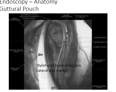
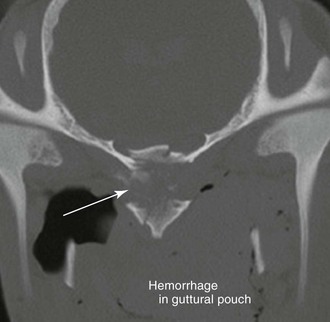
Gutteral pouch injury. Guttural pouch hemorrhage can also be associated with rupture of the rectus capitis ventralis and longus capitus muscles i e. It is caused by a fungus that infects the lining of the guttural pouch usually on the roof of the guttural pouch. Fungal plaques form within the guttural pouches most commonly along the walls of the major blood vessels internal carotid external carotid and maxillary arteries figure 3. Guttural pouch mycosis is a fungal infection of one or both guttural pouches.
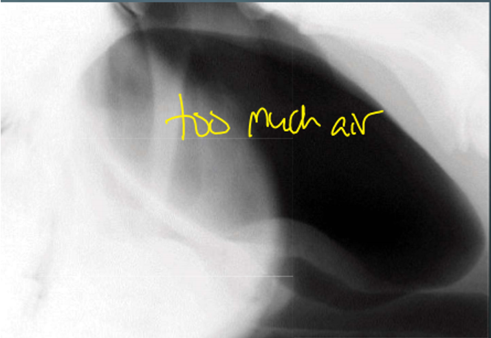
Guttural pouch mycosis is a rare but very serious disease in horses. They are lined with a thin membrane which separates them from nerves and and arteries. Tympany is usually unilateral but in some cases can affect both pouches. Guttural pouch mycosis is a fungal infection that affects horses.
The infection can cause some deep damage to the arteries and nerves. Guttural pouches are unique to few species of animals including the horse. The ventral straight muscles of the head or avulsion fracture of the basisphenoid bone where these muscles attach to it. The infection usually develops subsequent to a bacterial primarily streptococcus spp infection of the upper respiratory tract.
It is seen most often in young foals and is more common in females than in males. The physiological function if any of guttural pouches is unknown but recently it has been proposed that they might function during selective brain cooling sbc to maintain blood carried by the internal. Rearing over backwards and trauma to the base of the skull are often associated with rupture of these two muscles. Guttural pouch empyema is defined as the accumulation of purulent septic exudate in the guttural pouch.
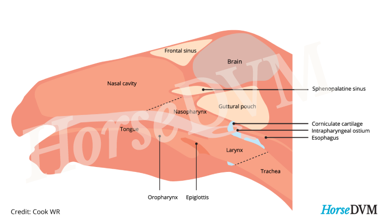
The most common causes of infection come from bacteria and fungi present within the pouch. The guttural pouch spans from the inside surface of the eardrum down to the pharynx the opening behind the mouth where the oral cavity and nasal passages meet. Redwings vet nic de brauwere talks us through the process as he scopes a horse s guttural pouch and takes sampl. Clinical signs include intermittent purulent nasal discharge painful swelling in the parotid area and in severe cases stiff head carriage and stertorous breathing.
These structures are large air filled sacs positioned on either side of the neck below the ear of the horse. The pouch wall is traversed by the internal carotid artery. What s involved in guttural pouch endoscopy.

